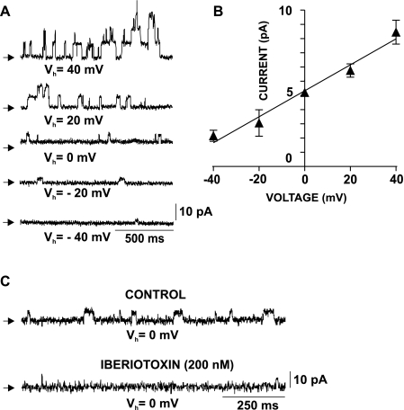
Fig. 7.
Single BK channel recordings in freshly isolated human DSM cells. A: original recordings showing a series of single BK channel openings at Vh from −40 to +40 mV in the whole cell configuration. The single BK channel amplitude was voltage dependent; increasing Vh toward positive voltages increased the single channel amplitude. Arrows indicate the closed channel state. B: current-voltage relationship for the single BK channel amplitude in freshly isolated human DSM cells. Values are means ± SE (n=11; N=10). C: original recordings showing the inhibitory effect of iberiotoxin on single BK channel currents at Vh=0 mV. Shown are current traces before (control) and 10 min after application of 200 nM iberiotoxin (n=5; N=5; P < 0.05). Ryanodine (30 μM), thapsigargin (100 nM), and nifedipine (1 μM) were present throughout the experiments to eliminate all Ca2+ sources for BK channel activation, hence TBKC.

