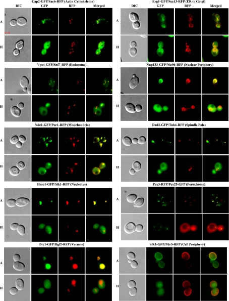
Fig. 5.
DIC, GFP, red fluorescent protein (RFP), and merged fluorescent images of cells expressing a hypoxia-redistributed and an unaffected protein in various organelles. Cells expressing a pair of GFP- and RFP-tagged proteins were grown in air (A) or under hypoxia (H), and the images were captured and are shown here. Except for the proteins showing a peroxisome location in air, the hypoxia-redistributed proteins were tagged with GFP, while the ones unaffected by hypoxia were tagged with RFP. For peroxisome, the RFP-tagged protein was affected by hypoxia, while the GFP-tagged protein was not. The scale bar represents 2 μm. The cellular locations of proteins in normoxic cells are also indicated at top.

