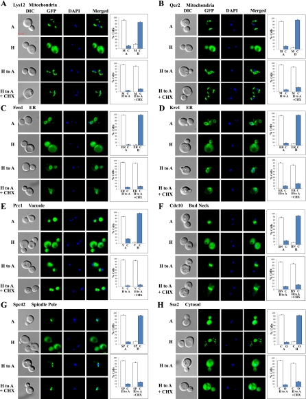
Fig. 8.
Examples of images of cells expressing redistributed nonnuclear proteins that recovered their normoxic nuclear locations in response to increased oxygen levels even in the presence of cycloheximide. Cells expressing GFP-tagged mitochondrial proteins Lys12 (A) and Qcr2 (B), ER proteins Fen1 (C) and Kre1 (D), vacuolar protein Prc1 (E), bud neck protein Cdc10 (F), spindle pole protein Spc42 (G), and cytosolic protein Ssa2 (H), respectively, were grown under hypoxic conditions and then shifted to normoxic conditions for 2 h in the presence or absence of cycloheximide. The images of cells grown under different conditions were captured. A, images of cells grown in air; H, images of cells grown in a hypoxia chamber; H to A, images of cells grown in a hypoxia chamber and then shifted to air for 2 h; H to A + CHX, images of cells grown in a hypoxia chamber and then shifted to air for 2 h in the presence of cycloheximide. The number of cells showing GFP-tagged proteins in the cytosol (C), mitochondria (M), vacuole (V), ER, bud neck (BN), spindle pole (SP), or other cellular locations (O) was counted and the percentage was calculated and is plotted. The scale bar represents 2 μm.

