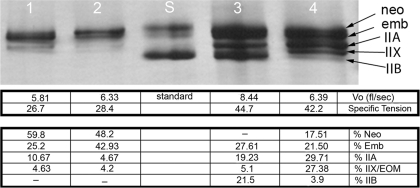
Fig. 3.
Representative silver-stained electrophoresis gel showing separation of myosin heavy chain (MyHC) isoforms obtained from single myofibers after force measurements had been obtained. The corresponding Vo, specific tension (mN/cm2), and percent composition of MyHC isoforms in each fiber are beneath each gel lane.

