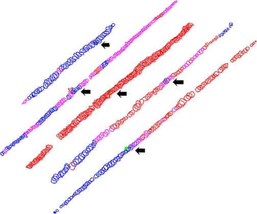
Fig. 8.
Reconstruction of 5 myofibers from serial sections immunostained for neonatal MyHC (Vector). Blue indicates regions that were negative, pink indicates regions that were stained at an intermediate level, and red indicates regions that were strongly positive. The green dot represents the section where the reconstructions were started. The reconstructed myofibers averaged 1.296 to 2.45 mm in length.

