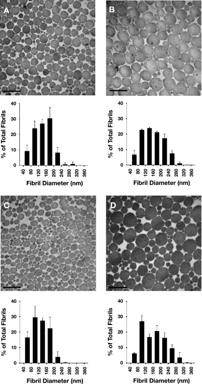
Fig. 4.
Representative electron micrographs and diameter distributions of collagen fibrils within distal (A and C) and proximal (B and D) regions of TA tendons of adult (A and B) and old (C and D) mice. In both age groups, diameters of fibrils tended to be larger in the proximal than distal region. Original magnification, ×64,000. Scale bar, 300 nm.

