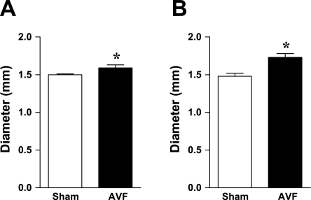
Fig. 3.
Diameter of the aorta and pulmonary artery in mice with AVF and in sham mice 2 wk after the creation of the AVF. Measurements of the root diameter of the aorta (A) and the pulmonary artery (B) were performed using echocardiography as described in materials and methods; n = 9 and n = 8 in sham and AVF groups, respectively. *P < 0.05 vs. sham.

