
Figure 1.
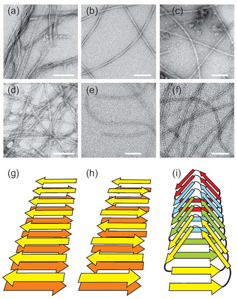
Negatively-stained TEM images of amyloid fibrils. (a) “Striated ribbon” Aβ1-40 fibrils. (b) “Twisted pair” Aβ1-40 fibrils. (c) Brain-seeded Aβ1-40 fibrils. (d) Amylin fibrils. (e) Sup35NM fibrils. (f) HET-s218-289 fibrils. White scale bars are 100 nm. (g,h,i) Schematic representations of cross-β structures formed by parallel β-sheets, antiparallel β-sheets, and a β-helix, respectively.