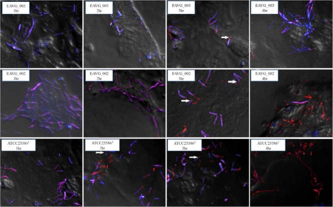
Fig. 2.
Time-dependent invasion of LS 174T cells by EAVG_002, EAVG_003, or ATCC 25586T. Bacterial cells were differentially stained using fluorescently labeled polyclonal antibodies, and merged images of fluorescent bacteria were deconvolved and overlaid on a snapshot phase-contrast image of the host cells. Internalized bacteria are stained orange, while those external to the host cell are stained purple. Arrows indicate examples of bacterial cells undergoing end-on invasion by the zipper mechanism; i.e., these cells are shown half internalized. All studied F. nucleatum isolates were invasive, but EAVG_003 is only minimally invasive. Differences in cell morphology between strains have been previously reported (37).

