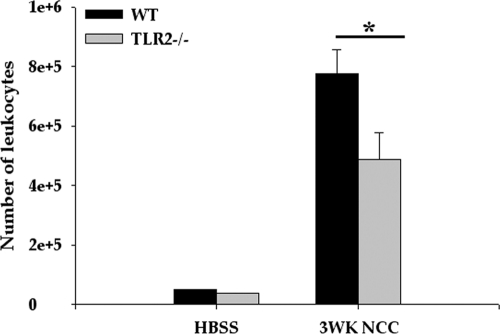
Fig. 4.
TLR2−/− mice exhibit reduced numbers of infiltrating immune cells in CNS. WT and TLR2−/− mice were i.c. infected with HBSS (mock control) or M. corti and were sacrificed at 3 weeks p.i. Infiltrating immune cells were isolated by Ficoll Hypaque density gradient as described in Materials and Methods. Total numbers of viable immune cells in mock-infected control and parasite-infected brains of both WT and TLR2−/− mice were counted by trypan blue exclusion staining (n = 4). A significant difference in numbers of immune cells recovered from TLR2−/− versus WT animals is indicated by an asterisk (P < 0.005).

