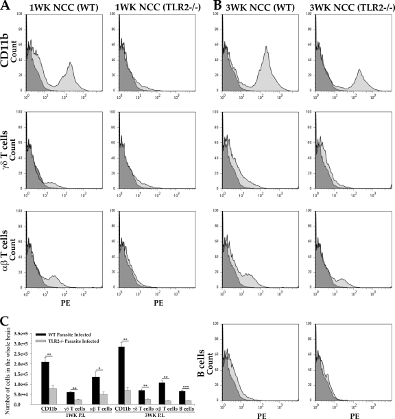
Fig. 5.
TLR2−/− mice exhibit reduced numbers of various infiltrating immune cells in CNS. WT and TLR2−/− mice were i.c. infected with M. corti and were sacrificed at 1 week and 3 weeks p.i. Infiltrating immune cells were isolated by Ficoll Hypaque density gradient as described in Materials and Methods. Total numbers of viable immune cells in parasite-infected brains of WT and TLR2−/− mice were counted by trypan blue staining. (A) In each experiment, brain leukocytes from 1-week parasite-infected WT and TLR2−/− mice were isolated from 3 mice and pooled together. The percentages of immune cell types that were macrophages/microglia (CD11b+), γδ T cells (T-cell receptor [TCR] δ2 chain-positive), αβ T cells (TCR β chain-positive), and B cells (CD19+) were quantified by FACS analysis. A representative of three independent experiments is shown. (B) Brain leukocytes from 3-week parasite-infected WT and TLR2−/− mice were isolated from 3 mice and pooled together. FACS analysis shows a reduced number of macrophages/microglia (CD11b+), γδ T cells (TCR δ2 chain-positive), and αβ T cells (TCR β chain-positive) in TLR2−/− mice. A representative of three independent experiments is shown. (C) Results obtained from FACS analyses are expressed as the mean ± standard error of the mean from three independent experiments. Significant differences in numbers of individual immune cells recovered from TLR2−/− versus WT animals are denoted by asterisks (*, P < 0.05; **, P < 0.005; ***, P < 0.005).

