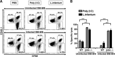
Fig. 4.
Leishmania-infected macrophages are resistant to NK cell lysis in vivo. WT B6 mice (CD45.1−) were treated i.p. with PBS, poly(I:C) (50 μg), or L. infantum promastigotes (107 parasites). At 18 to 20 h posttreatment or postinfection, 2.5 × 10⁵ CFSE-labeled uninfected or infected BM-Mφ (CFSEhi CD45.1+, L. infantum promastigote/cell ratio for infection = 7:1) from congenic B6 PTPRC mice were injected i.p. along with an equal number of uninfected or infected MHC class I-deficient (β2m−/−) BM-Mφ (CFSEhi CD45.1−, parasite/cell ratio for infection = 7:1) and uninfected WT splenocytes (CFSElo CD45.1+). (A) The percentage of CFSE-labeled cells recovered from the peritoneal cavity at 14 to 16 h posttransfer was analyzed by flow cytometry. After gating on CFSE-positive cells, the transferred populations were distinguished on the basis of their differential intensity of CFSE fluorescence (high or low) and expression of CD45.1 (positive or negative). (B) The bar graph represents the mean (±SEM) of percent specific lysis obtained from four independent experiments with four mice per group as described in Materials and Methods. ***, P < 0.0001 (Mann-Whitney test).

