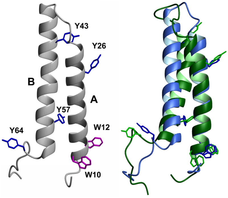
Figure 1. Structure of bacteriorhodopsin peptide AB.

Left side: Amino acids 5–71 from the crystal structure of bacteriorhodopsin (Protein Data Bank 1C3W), comprising transmembrane helices A and B. Tryptophan (W) and tyrosine (Y) residues are indicated. Right side: Superposition of peptide AB structures from final frames of MD simulations in POPC belayed (blue) or SDS micelle (green). Figure generated using Molmol [41].
