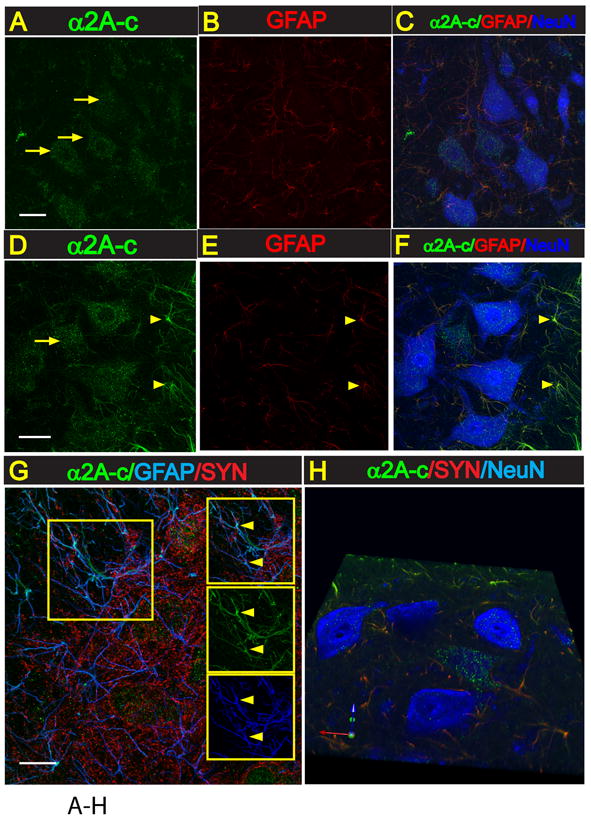
Fig. 4. Increased expression of α2A-c receptor in activated astrocytes in lumbar spinal cord in rats with increased stretch reflex activity.

A-F: Transverse lumbar (L3) spinal cord section taken from naive non-ischemic animal (A-C) or from an animal with spinal ischemic spasticity and triple-stained with α2A-c, GFAP and NeuN antibody. In control naive animals a punctuate-like α2A-c immunoreactivity in virtually all neurons including small and medium-sized interneurons and α-motoneurons can be seen (A). Co-staining of the same sections with GFAP antibody showed only weak expression of α2A-c in astrocytes (A, B). In contrast to control animals, in rats with ischemic injury a clear increase in α2A-c expression in activated astrocytes localized at the peri-ischemic regions in the vicinity of persisting α-motoneurons can be identified (D-F; yellow arrows).
G: Confocal images taken from transverse spinal cord lumbar section from an animal with spinal ischemic injury and stained with α2A-c, GFAP and synaptophysin antibody. A clear colocalization of α2A-c and GFAP expression in astrocyte processes can be seen (G-yellow arrows).
H: 3-D image of α2A-c overexpressing astrocytes (green) in the vicinity of lumbar α-motoneurons (blue).
