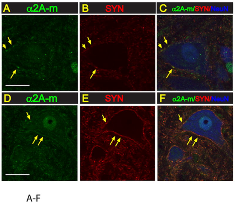
Fig. 5. Expression of α2A-m receptor in lumbar spinal cord interneurons and α-motoneurons.

A-F:Transverse lumbar (L3) spinal cord section taken from naive non-ischemic animal (A-C) or from an animal with spinal ischemic spasticity and triple-stained with α2A-m, synaptophysin and NeuN antibody. In both control and spinal ischemic animals a punctate-like α2A-m immunoreactivity can readily be identified on α-motoneuronal membranes (yellow arrows). A spatial colocalization of α2A-m punctata and synaptophysin expression can also be seen (yellow arrows).
