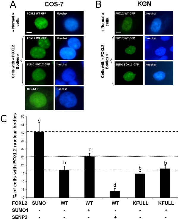
Figure 3. SUMOylation promotes FOXL2 recruitment to subnuclear structures.
A and B) Presence of nuclear structures displaying high FOXL2-GFP concentration in a climate of high SUMOylation. Micrographs of COS-7 cells (A) and KGN cells (B) expressing FOXL2-GFP, the tripartite SUMO-FOXL2-GFP fusion or NLS-GFP, as indicated. Left column: GFP. Right column: Hoechst 33342 DNA staining. First line shows a representative “normal” cell, where FOXL2 displays its most common localisation, superimposable to DNA. Second and third lanes show representative cells with “FOXL2 nuclear bodies” where FOXL2 is enriched in subnuclear structures clearly distinct from chromatin structures. Fourth lane (only in A) shows a typical cell stained with nuclear localised GFP. Scale bar: 5 µm. Scale bar is valid for all micrographs. (C) Quantification of cells presenting subnuclear structures in COS-7 expressing SUMO-FOXL2-GFP, FOXL2-WT-GFP or FOXL2-KFULL-GFP with or without overexpression of SUMO1 or SENP2. At least 300 GFP-positive cells were scanned per condition (in groups of 50 to estimate the standard deviations). Error bar: SEM. Letters a, b, c, d refer to statistical categories in a Student's t-test. Conditions with different letters are statistically different with p<0.05.

