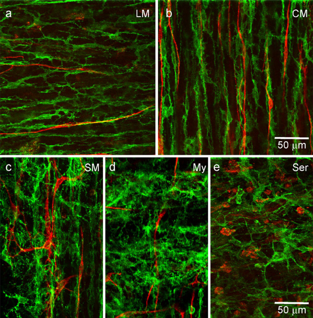
Fig. 7.
PDGFRα+ cells are closely associated with ICC-IM in WT mouse IAS. Dual-labeling of PDGFRα+ cells (green) and ICC (red) in whole-mount preparations of the IAS of the WT mouse. ICC-IM are closely associated with PDGFRα+-IM in the LM (a) and CM (b) layers. In contrast, ICC located near the submucosal (SM, c) and myenteric (My, d) surfaces appear to have little relationship to PDGFRα+ cells. Round KIT+ cells seen within the serosa (Ser, e) are likely to be mast cells and do not appear to have a close association with PDGFRα+ cells. Optical section thicknesses: a 2.5 µm, b 3 µm, c 5.5 µm, d 2.5 µm, e 3.5 µm

