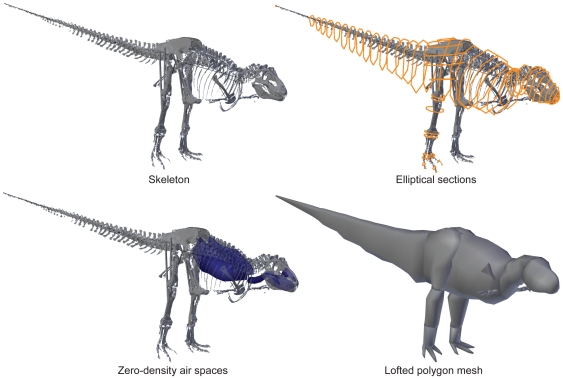
Figure 1. Modelling procedure, showing the Carnegie specimen.
From left to right, top to bottom these show the scanned, reconstructed, and straightened skeleton; the skeleton with elliptical hoops that define fleshy boundaries; the air spaces representing pharynx, sinuses, lungs and other airways including air sacs; and the final meshed reconstruction used for mass and COM estimates.

