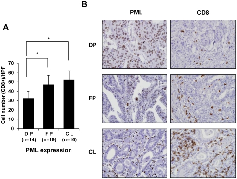
Figure 1. Promyelocytic Leukemia (PML) Protein Expression is Inversely Correlated with the Extent of Infiltration of CD8+ T-cells in Advanced Gastric Carcinoma Tissues.
(A) Immunohistochemical staining for PML protein and CD8 was performed for 49 cases of stage IV advanced gastric carcinomas. Immunopositivity for PML protein was categorized as diffuse positivity (DP; nuclear immunoreactivity in ≥50% of tumor cells, n = 14), focal positivity (FP; in ≥10% but <50%, n = 19), or complete loss (CL; in <10%, n = 16). The number of CD8 immunopositive cells in one high power field (HPF, x40) of 0.25 mm2 for five randomly selected tumor infiltrative borders were counted for each specimen, followed by calculation of the mean and SD. (B) Representative images showing PML protein expression and CD8+ T-cell infiltration in advanced gastric carcinoma tissues. Bars, SD. *P<0.05.

