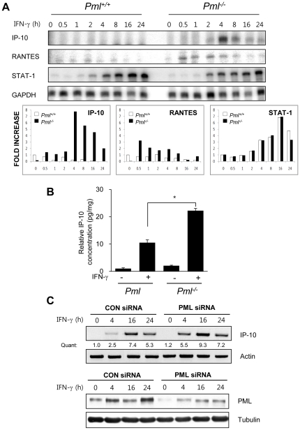
Figure 3. Expression Levels of IFN-γ-inducible protein-10 (IP-10) are Enhanced in Pml .
−/− MEFs compared to Pml+/+ wild-type cells. (A) RNA from Pml+/+ and Pml −/− MEFs treated with IFN-γ (10 ng/ml) for 0-24 h was subjected to ribonuclease protection assay (RPA). Quantification of IP-10, RANTES and STAT-1 mRNA (FOLD INCREASE) was calculated by dividing the densitometric value of each lane by the corresponding GAPDH value. (B) Pml+/+ and Pml −/− MEFs cells were incubated in the absence or presence of IFN-γ (10 ng/ml) for 12 h. IP-10 protein from the culture supernatants were analyzed by ELISA and normalized by the cell protein concentration. The relative concentration was calculated as the normalized amount divided by the normalized amount of untreated Pml −/− MEFs cells. (C) NIH3T3 fibroblasts were transiently transfected with either Pml siRNA or control siRNA. Two days after transfection, cells were treated with or without IFN-γ for 0-24 h, and analyzed by immunoblotting for detection of PML protein expression and RT-PCR for IP-10 mRNA. Quantification (Quant) of IP-10 mRNA was calculated by dividing the densitometric value of each lane by the corresponding actin mRNA value. Data shown are representative of at least three experiments. Bars, SD. *P<0.01.

