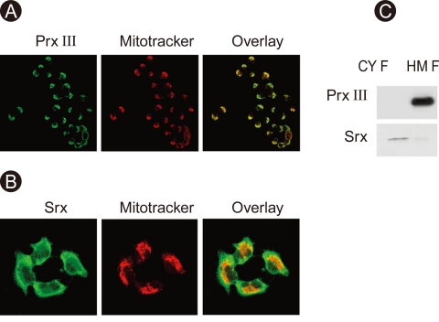
Figure 1.
Expression and cellular localization of peroxir-edoxin (prx) III and sulfiredoxin (srx). A549 cells were stained for Prx III (FITC), Srx (FITC), and mitochondrial fraction (Mitotracker) and then examined by confocal microscopy (A, B). A549 cells were subjected to subcellular fractionation to yield mitochondria-enriched heavy membrane (HM) and cytosolic fractions (CYF). Cell lysates were prepared and subjected to immunoblot analysis with antibodies to either Prx III or Srx (C).

