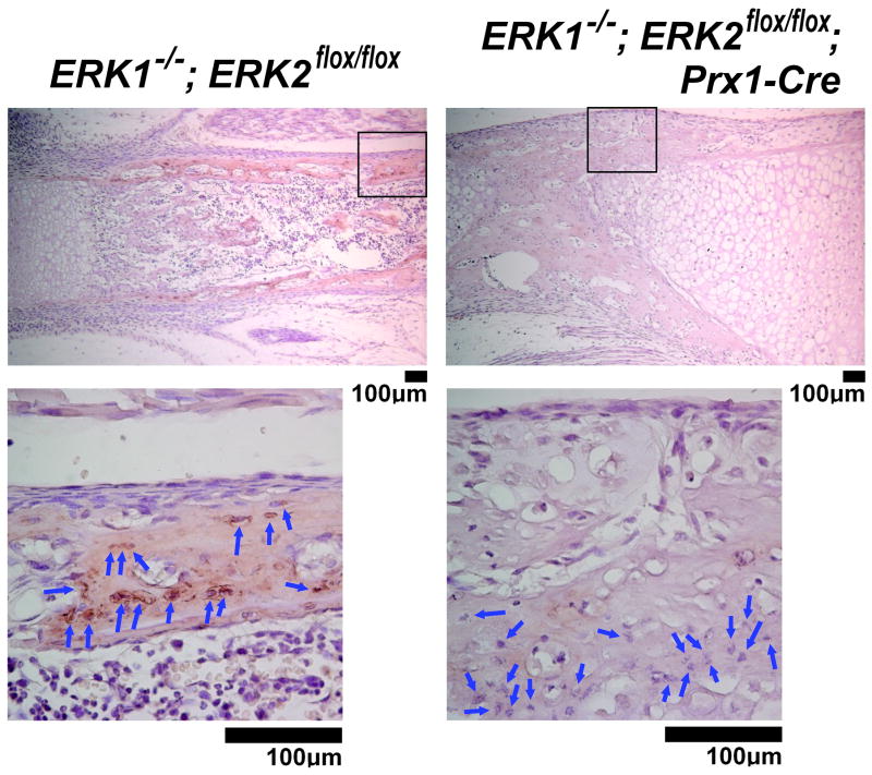
Fig. 12.
Immunohistochemical analysis for Dmp1 protein expression in the tibiae of ERK1−/−; ERK2flox/flox; Prx1-Cre mice at postnatal day 0. The staining for Dmp1 was remarkably reduced in osteocytes (blue arrows) of ERK1−/−; ERK2flox/flox; Prx1-Cre mice, whereas control littermate ERK1−/−; ERK2flox/flox mice showed intense staining in osteocytes and their surrounding matrices. The boxed area in the upper panel is magnified in the bottom panel.

