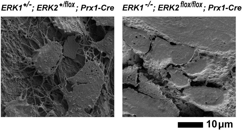
Fig. 13.
Scanning electron microscopy images of the acid-etched, resin-casted osteocyte lacunar-canalicular system in the tibia at postnatal day 0. While control ERK1+/−; ERK2+/flox; Prx1-Cre mice show osteocytes with characteristic dendritic processes and organized canaliculi (left panel), osteocytes in ERK1−/−; ERK2flox/flox; Prx1-Cre mice lack dendritic processes, and no canalicular system is observed.

