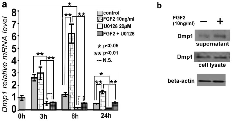
Fig. 5.
FGF2 increases Dmp1 mRNA and protein levels in MLO-Y4 cells. (a) Time-dependent increase of Dmp1 mRNA after FGF2 treatment. The cells were treated with 10 ng/ml FGF2 in the presence or absence of 20 μM U0126. RNA was isolated at 3, 8 and 24 h after FGF2 treatment. Dmp1 mRNA levels were measured by real-time PCR in triplicate. Data represent means ± standard deviations. (b) FGF2 increases Dmp1 protein levels in the culture supernatant of MLO-Y4 cells, while Dmp1 protein levels in total cell lysates remain unchanged. MLO-Y4 cells were treated with 10 ng/ml FGF2 in the absence of serum for 21 h. Culture supernatant was concentrated using centrifugal filter units and subjected to Western blot analysis. The bottom panel shows β-actin protein levels in total cell lysates as a loading control. The figure presents data from one of four experiments with similar results. (N.S.) Not significant.

