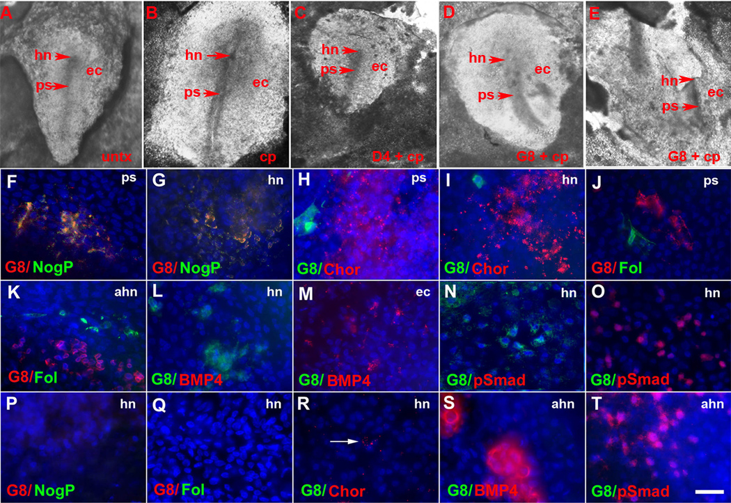
Figure 4. Effects of Myo/Nog cell ablation on the expression of regulators of BMP signaling in gastrulating embryos.
Untreated embryos (untx) and embryos incubated with complement (cp) only or D4 and complement at stage X–XII displayed a normal primitive streak (ps) by stage 3–4 (A–C). G8 ablated embryos had primitive streaks of abnormal morphology and/or positioning (D, E). Stage 3–4 embryos were double labeled with antibodies. The photographs of the embryos in A and D demonstrate the regions shown in the photomicrographs of F–O and P–T, respectively. The colors of the fluorescent tags are indicated in each panel. Overlap of red and green appears yellow in merged images. In untreated embryos, G8+/noggin+ (NogP) cells were present in the primitive streak (ps) (F) and Hensen’s node (hn) (G). Chordin (Chor) was found in the primitive streak (H) and Hensen’s node (I). Staining for follistatin (Fol) was observed in the anterior primitive streak (J) and above Hensen’s node (ahn) (K). BMP4 (L) and p-Smad1/5/8 (N) were absent in Hensen’s node but present in the lateral ectoderm (ec) (M and O). Staining for chordin, follistatin, BMP4 and p-Smad1/5/8 did not overlap with G8 (H–O). Neither G8, noggin nor follistatin was detected in Hensen’s node in embryos ablated with G8 and complement (P and Q). Staining for chordin was weaker in Hensen’s node of G8 ablated embryos (R) compared to untreated embryos (I). BMP4 (S) and p-Smad1/5/8 (T) were present above Hensen’s node in G8 ablated embryos. Bar, 135 µm in A–E and 9 µm in F–T.

