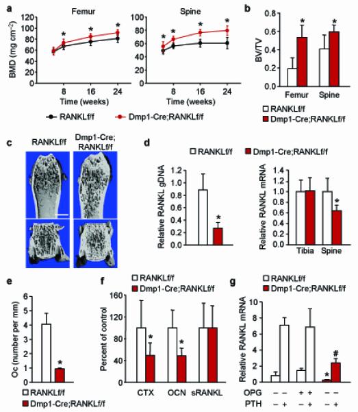
Figure 3. Deletion of RANKL from Dmp1-Cre expressing cells reduces bone remodeling.

(a) Serial BMD of Dmp1-Cre;RANKLf/f (n = 14) and RANKLf/f (n = 19) littermates. *P < 0.05 using Student’s t-test comparing the two genotypes at a given age. (b) Cancellous bone volume in the distal femur or in L4 vertebra of 6-month-old Dmp1-Cre;RANKLf/f (n = 11) and RANKLf/f (n = 7) littermates. *P < 0.05 using Student’s t-test. (c) Representative μCT images of the distal femur and L4 vertebra of 6-month-old Dmp1-Cre;RANKLf/f and RANKLf/f littermates. Scale bar, 1 mm. (d) Left, quantitative PCR of loxP-flanked RANKL genomic DNA using genomic DNA isolated from collagenase-digested femoral cortical bone of 6-month-old Dmp1-Cre;RANKLf/f (n = 11) and RANKLf/f (n = 7) littermates. Right, quantitative RT-PCR for RANKL mRNA in tibia and L5 vertebra of the same mice as in the left panel. *P < 0.05 using Student’s t-test. (e) Osteoclast number per mm bone surface in cancellous bone of the distal femur of 6-month-old Dmp1-Cre;RANKLf/f (n = 4) and RANKLf/f (n = 4) littermates. *P < 0.05 using Student’s t-test. (f) Carboxy-terminal crosslinked telopeptide of type I collagen (CTX), osteocalcin (Ocn), or soluble RANKL in the blood plasma of 6-month-old Dmp1-Cre;RANKLf/f (n = 9) and RANKLf/f (n = 8) littermates. *P < 0.05 using Student’s t-test. (g) RANKL mRNA levels in tibial cortical bone of 6-month-old Dmp1-Cre;RANKLf/f and RANKLf/f littermates, pretreated with vehicle or OPG and then injected with vehicle or PTH(1-34) (n = 6 to 8 per group). *P < 0.05 versus RANKLf/f mice pretreated with vehicle or OPG and then injected with vehicle using 2-way ANOVA. #P < 0.05 versus RANKLf/f mice pretreated with vehicle or OPG and then injected with PTH using 2-way ANOVA. All values include data from both sexes.
