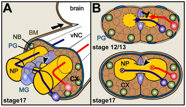
Figure 2.
Cellular constellations in the embryonic ventral nerved cord of Drosophila
A) The mature ventral nerve cord (vNC; equivalent to spinal cord; arrow points anterior) at embryonic stage 17 (Campos-Ortega and Hartenstein, 1997) is ensheathed by perineurial and subperineurial glia cells (blue line; PG) (Ito et al., 1995) covered by BM (brown line). Quiescent neural precursor cells (neuroblasts, NB) and neuronal cell bodies lie in the cortex (CX) from where axons project straight into the neuropile (NP; the synaptic compartment, which is itself ensheathed by interface glia; not shown) where they constitute ascending and descending pathways, dendrites and synapses (Sánchez-Soriano et al., 2007). Contralateral neurons (blue) project across the midline through two segmental commissures (arrow head) enwrapped by midline glia (MG). B) The lower image shows a frontal cross section of the vNC in A, the upper image the same section at an earlier stage when axonal growth takes place. Slit is released from midline glia cells and repels (black T-bar) growth cones of Robo-presenting neurons (red). At this early stage, the BM (dashed brown line) and PG sheath are incomplete, and fat body/hemocyte-derived ECM proteins like Tiggrin, Cg25C or Laminin A are enriched in the NP.

