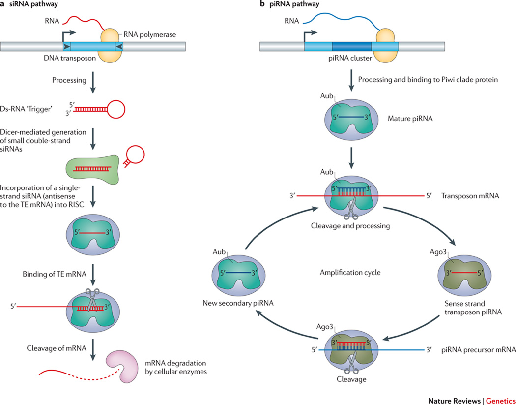
Figure 3. The degradation of transposon mRNA by RNAi.
A. The siRNA pathway: Double-stranded “trigger” RNAs (hairpin), derived from the inverted terminal repeats of a DNA transposon in the case illustrated, are processed and then cleaved into 21 to 24 nt siRNAs by the dicer family of proteins (light green amorphous shape). A single-strand siRNA (short red line) complementary to the transposon mRNA is selectively incorporated into the Argonaute containing RISC complex (blue amorphous shape with short red line). The siRNA directs RISC to complementary sequences in transposon mRNA (long red line), leading to its post-transcriptional destruction. The figure was drawn based on concepts presented in the following reviews147, 148, 152. B. The piRNA pathway: A primary piRNA transcript (wavy blue line) generated from a piRNA cluster (blue rectangle) that contains sequences derived from TEs (darker blue rectangle inset) is processed into mature 24–35 nt piRNAs (small blue line). Binding of the mature piRNA by the Piwi/Aubergine protein (color?…make different from Ago3 and panel A) allows it to be directed to complementary sequences in TE mRNA (red line). Endonucleolytic cleavage (scissor) 10 nt from the 5’ end of the small RNA and 3’ processing liberates a secondary sense strand transposon piRNA (red line), which associates with the Argonaute 3 protein (color?…make different from Aubergine and panel A). The binding of this complex to complementary sequences in the original precursor piRNA, followed by endonucleolytic cleavage and 3’ processing liberates, generates a secondary antisense piRNA that can be directed to TE mRNA. This reiterative cycle (e.g., a “ping-pong” cycle) can lead to the destruction of transposon mRNA in the germ line. The piRNA model was redrawn based on concepts and models presented in the following papers30, 151, 157, and for the example illustrated is from Drosophila; however, a similar pathway likely operates in mammalian cells (see text for details).

