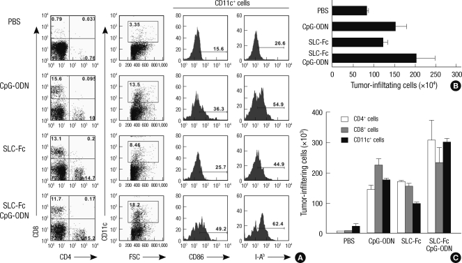
Fig. 4.
Recruitment of T cells and DCs inside the tumor. B16F10 melanoma cells (2 × 10⁵) were inoculated in the shaved left flank of B6 mice. When the tumor volume reached approximately 150 µL, mice were received with PBS, 10 µg CpG-ODN (twice), 3 µg SLC-Fc (twice), or 10 µg CpG-ODN plus 3 µg SLC-Fc (twice). Eleven days after intratumoral administration of CpG-ODN and/or SLC-Fc, tumors were removed. Infiltrating cells in the tumor mass were obtained by treatment with collagenase D (100 µg/ea) for 25 min at 37℃ and stained with anti-CD4, anti-CD8, anti-CD11c, anti-CD86, and anti-I-Ab (Y3P) antibodies. (A) Populations of the infiltrating cells in the tumor mass were analyzed by flow cytometry. (B) Total numbers of the infiltrating cells in the tumor mass were counted. (C) Each population of the infiltrating cells in the tumor mass was calculated from data based on flow cytometry analysis. Data represent means±SD of two independent determinations with n = 4 mice/group.

