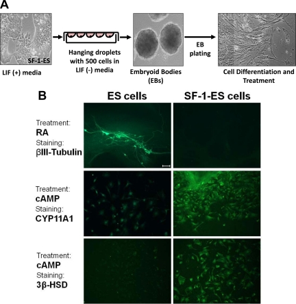
Fig. 3.
Differentiation of SF-1-ES cells after the formation of EBs. A, Schematic illustration of the protocol for differentiation of cells through EB formation. B, ES cells and SF-1-ES cells were differentiated as EBs and then treated with RA for neuronal differentiation or with cAMP and stained with neuronal (βIII-tubulin) or with steroidogenic markers (CYP11A1; 3β-HSD). Bar, 100 μm.

