
Figure 5.
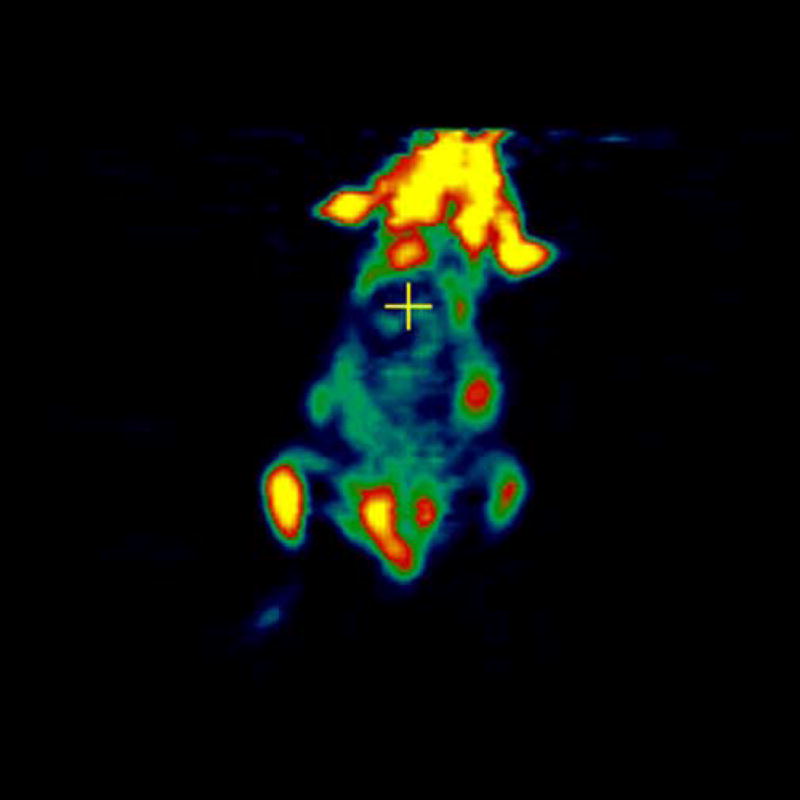
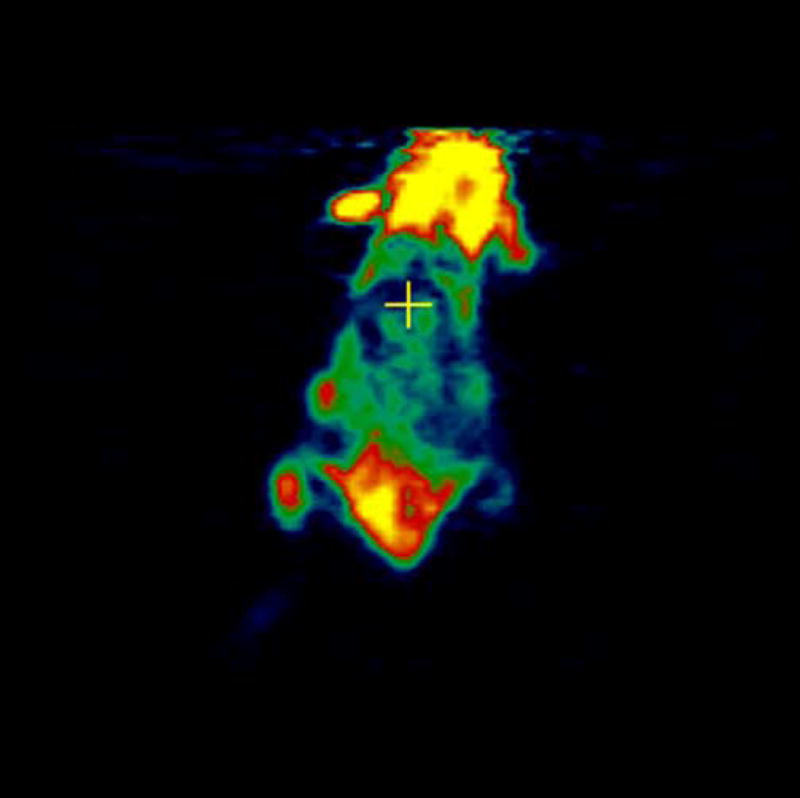
microPET images showing immunolocalization of 18F-FDG in mice with 4T1 and CMS4 tumors. 5a. the planar slice which showed maximum 4T1 localization is shown. 5b. the planar slice which showed maximum CMS4 localization is shown. Tumors were not on the same plane, therefore 2 images were used. Before scanning, the mice were anesthetized with O2/isoflurane (1%-3% isoflurane) and imaged in the prone position in the gantry of the microPET scanner and the animal was scanned for 15 minutes for [18F]-FDG.