
Figure 5.
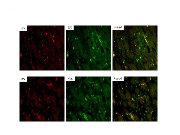
Dual immunofluorescent staining for cells expressing αv in ischemic limb sections. Sites of αv expression were shown to be mainly endothelial cells based on colocalization of αv (Texas Red) with FVIII (green, fluorescein isothiocyanate) in the merged image. Colocalization of αv with macrophages (Mac-3, fluorescein isothiocyanate) was also seen in the merged image. Areas in yellow represent colocalization. EC, endothelial cells. (Magnification ×200).