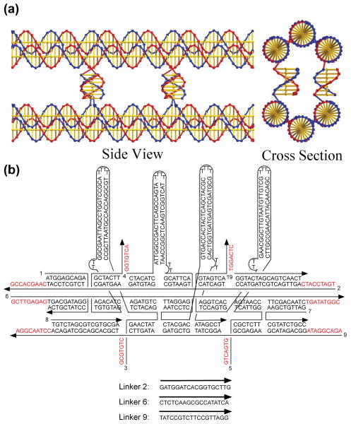
Figure 1. DNA tile sequences and structures.
(a) The P6HB Motif. This representation24 shows the way in which two BTX domains are paired by four lateral connections to form the P6HB motif. The cross section view shows two of the four helices that are formed by the lateral cohesive interactions. The interactions at the rear are eclipsed in this projection. (b) The Sequence and Structure of the B′ BTX Tile. Four helical domains, hairpins, are shown attached perpendicular to the BTX motif, so that they will create a topographic feature that can be detected in the atomic force microscope (AFM). Other tiles are shown in the Supplementary Information (S1).

