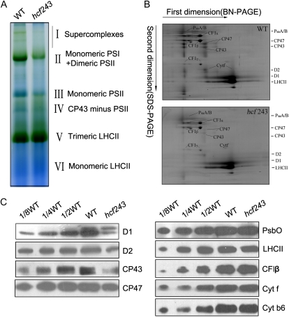
Figure 4.
Analysis of thylakoid proteins from wild-type (WT) and hcf243 plants. A, Two-dimensional separation of protein complexes in the thylakoid membranes. BN-PAGE-separated thylakoid proteins in a single lane from a BN gel were separated in a second dimension by 15% SDS-urea-PAGE and stained with Coomassie blue. B, BN gel analysis of thylakoid membrane (10 μg of chlorophyll) from wild-type and hcf243 plants was solubilized and separated by BN gel electrophoresis. The positions of protein complexes were identified with appropriate antibodies (Guo et al., 2005). C, Immunoblot analysis of thylakoid proteins from the wild type and hcf243. The thylakoid membrane proteins were separated by SDS-urea-PAGE, and the blots were probed with specific anti-D1, anti-D2, anti-CP47, anti-CP43, anti-CF1β, anti-LHCII, anti-PsbO, anti-Cytf, and anti-Cytb6 antibodies.

