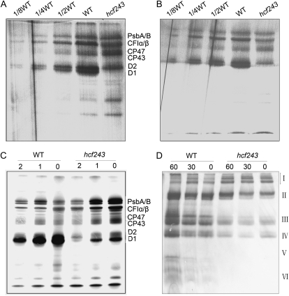
Figure 6.
In vivo [35S]Met labeling of thylakoid proteins from wild-type (WT) and hcf243 plants. A and B, Pulse labeling of thylakoid membrane proteins in 14-d-old young seedlings (A) and 4-week-old plants (B). After pulse labeling young Arabidopsis seedlings in the presence of cycloheximide for 20 min, thylakoid membranes were isolated, and the proteins were separated by SDS-urea-PAGE and visualized autoradiographically. C, Pulse and chase labeling of thylakoid membrane proteins. After 30 min of pulse labeling in 14-d-old young seedlings in the presence of cycloheximide followed by 1 or 2 h of chase with cold Met, thylakoid proteins were separated by SDS-urea-PAGE and visualized autoradiographically. D, BN -PAGE analysis of the incorporation of [35S]Met into thylakoid membrane protein complexes. A 15-min pulse in Arabidopsis young seedlings in the presence of cycloheximide was followed by a chase of cold unlabeled Met for 30 and 60 min. The thylakoid membranes were isolated and solubilized with DM, and the protein complexes were separated by BN-PAGE and visualized by autoradiography. Bands corresponding to various PSII assembly complexes of PSII supercomplexes (band I), monomeric PSI superimposed on the PSII dimer (band II), monomeric PSII (band III), CP43-free PSII monomer (band IV), reaction center (band V), and free proteins (band VI) are indicated at right.

