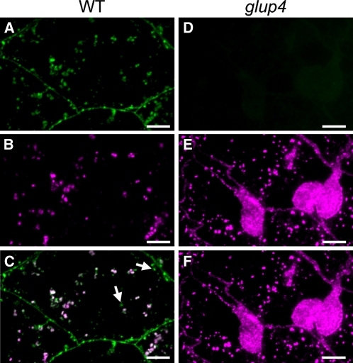
Figure 7.
Subcellular localization of Rab5a in 2-WAF developing wild-type and glup4 (EM956) seeds by immunofluorescence microscopy. A to C, The wild type (WT). D to F, glup4 (EM956). Secondary antibodies labeled with FITC (green) and rhodamine (magenta) were used to visualize the reaction of Rab5a in A and D and glutelin antibodies in B and E, respectively. C and F are the merged images of A/B and D/E, respectively. Arrows indicate the granules showing only Rab5a signal. Bars = 10 μm.

