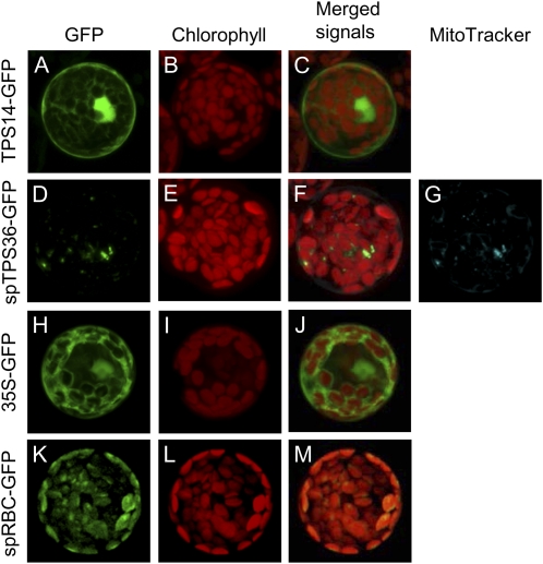
Figure 2.
Subcellular localization of TPS14 and TPS36. The complete open reading frame of TPS14 (TPS14-GFP) and the first 60 codons of TPS36 (tpTPS36-GFP) were fused to a downstream GFP and transiently expressed in Arabidopsis leaf protoplasts. GFP fluorescence indicates the location of each fusion protein; the location of the chloroplasts was determined by chlorophyll autofluorescence (shown in red), and the location of mitochondria was determined by the fluorescence of MitoTracker Red dye (shown in artificial blue color to distinguish it from chlorophyll autofluorescence). The column labeled “Merged Signals” provides a view of all fluorescent signals obtained for this sample. A to C, TPS14-GFP. D to G, tpTPS36-GFP. H to J, Expression of a nonfused GFP showing cytosolic (and nuclear) localization, used here for control. K to M, Expression of a Rubisco-transit peptide-GFP fusion gene (Nagegowda et al., 2008) showing plastid localization of the protein, used here for control.

