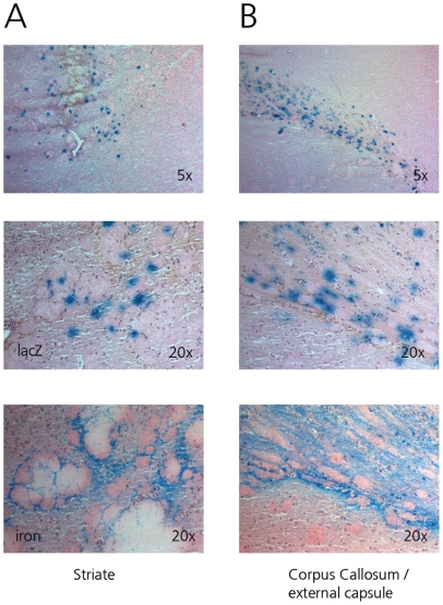
Figure 4. Determination of infected cells by nuclear morphology (top 2 rows, original magnification 5× and 20×, HE staining).
β-Gal staining shows glial cells are infected following infusion of AdCMVLacZ in the rat striate (A) or external capsule (B). No infected neurons were detected. No infected cells were detected in the overlying neocortex. SPIO distribution (iron staining) is similar to β-Galactosidase staining both in gray and white matter (bottom row, original magnification 20×).

