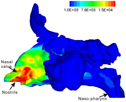
Figure 7. Contour plot of nasal mucosal heat flux (J/m2) for a subject that received a nasal/sinus CT scan immediately before testing.
A CFD model was created for this subject using the method described by Zhao et al. [16] in which nasal airflow and mucosal heat exchange are then simulated. The wall boundary condition at the mucosal surface is set similar to that described by Naftali et al. [14]: fully saturated with water vapor and at body temperature, with an unlimited supply of heat and water vapor.

