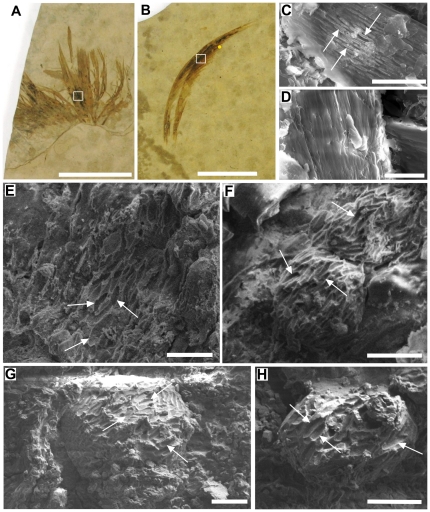
Figure 1. Visual and SEM images showing the presence of melanosomes in Gansus yumenensis and extant feathers.
Isolated feathers (A) MGSF318 and (B) MGSF317, with SEM images of the fractured surfaces of (C) an extant Marabou stork feather and (D) a White-naped Crane feather, and the dark areas of MGSF317 (E-F) and MGSF318 (G-H). Eumelanosomes present within the extant Marabou stork feather (C) and the elongate mouldic structures interpreted as eumelanosomes in the fossil feathers (E-H) are highlighted with white arrows. Scale bars represent 2 cm (A-B), 5 µm (C–D) and 2 µm (E–H). The fossil feather SEM images were taken from the areas indicated by the white boxes, and the yellow dot in (B) represents the approximate area where the FTIR map discussed below is taken from.

