Figure 1.

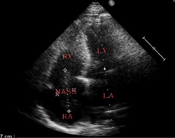
Apical four chamber view showing right atrium filled with a medium echogenic oval tumor mass by transthoracic two-dimensional echocardiography. RV: right ventricle, RA: right atrium, LV: left ventricle, LA: left atrium.

Apical four chamber view showing right atrium filled with a medium echogenic oval tumor mass by transthoracic two-dimensional echocardiography. RV: right ventricle, RA: right atrium, LV: left ventricle, LA: left atrium.