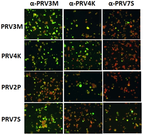
Figure 1. Immunofluorescence staining of four PRVs using human patient sera.
MDCK cells infected with PRV3M, PRV4K, PRV2P and PRV7S (presented in panels from top to bottom), respectively, were each probed with convalescent serum from patient infected with PRV3M, PRV4K and PRV7S (from left to right), respectively. All human sera were used at 1∶40 dilution.

