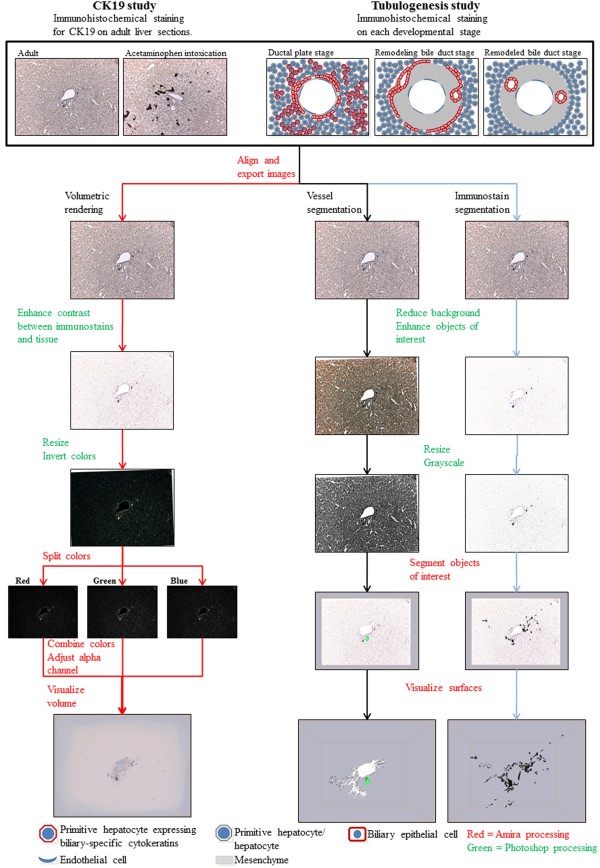
Figure 1.
Overview of the 3D reconstruction process. Serial formalin-fixed and paraffin-embedded liver sections were immunohistochemically stained and photographed. The images were aligned with Amira, exported and copied to three different stacks; one for the volumetric rendering and two for segmentation of the vessels and immunostains, respectively. With the purposes of enhancing the objects of interest the images were modified with Adobe Photoshop and resized. In Amira the color channels of the image stacks for the volumetric rendering were separated followed by their recombination and calculation of a new transparency channel. Visualization was completed through the use of the Voltex module. For the segmentation of the portal vessels and immunostains, each object of interest was labeled with Amiras built-in segmentation editor followed by their visualization with the SurfaceGen and SurfaceView modules.

