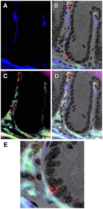
Figure 2. Double IHC for α-SMA and CD45 with Y chromosome paint.
A colonic crypt from a female Rag2−/− mouse transplanted 28 days earlier with male SWR BM. A. α-SMA was stained using two layer IHC with a Cy5-conjugated second layer, here false-coloured blue. B. CD45 was revealed using 3 layer IHC using Vector Red as chromogen for alkaline phosphatase-conjugated streptavidin, here revealed in fluorescent mode using the Cy3 filter, in combination with the brightfield greyscale image (with nuclei stained with haematoxylin), and overlaid with the α-SMA image from A. Note the close morphology of the CD45-positive cell to that of its neighbours. C. Subsequent Y chromosome paint (green nuclear spots, FITC filter) on the same section. Note background fluorescence revealing some tissue morphology. D. Overlay image of B and C showing the CD45 cell to be Y chromosome-positive, together with some stromal male cells. E. Magnified view of part of D. Original magnifications: A-D 200X, E 400X.

