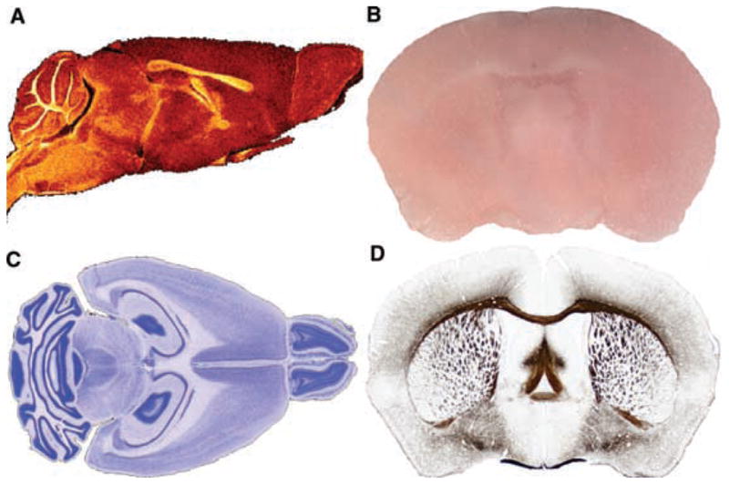
FIG. 1.

A digital atlas: orthogonal sections and high-resolution display. (A) Horizontal, (B) transverse (coronal), and (C) sagittal sections through two volumes, an MRM volume and a Nissl-stained volume from a d100 mouse, shown overlaid. Small, low-resolution thumbnails are for navigation, whereas, (D) a high-resolution view of the same data allows one to visualize both nuclei and white matter tracts.
