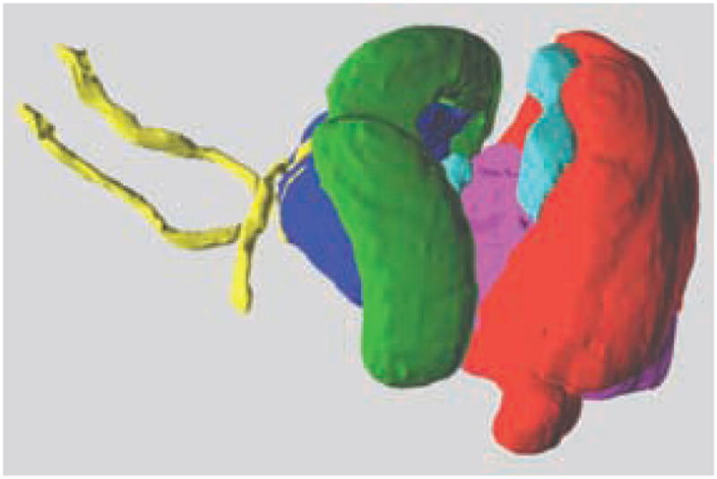
FIG. 2.

3D surfaces displayed in space. 3D structures were created by tessellating volumetric anatomical delineations. The outer surface of the brain and some deep structures have been rendered transparent to visualize the anterior commissure, the hippocampus, and the thalamus in the anterior part of the brain, and the cerebellum, inferior colliculi, and hindbrain in the posterior part.
