
FIG. 3.
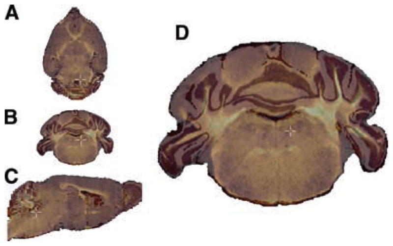
Multiple modalities and planes of section. Data are shown in several planes of section to demonstrate the inherently 3D nature of the atlas. (A) An MRM scan of a d100 mouse brain using a z-direction diffusion-weighted imaging protocol. (B) A transverse (coronal) section from a blockface imaging volume of a d100 mouse brain. (C) A horizontal section from a Nissl-stained volume of a d123 mouse brain. (D) A transverse (coronal) section from a myelin-stained volume of a d100 mouse brain.