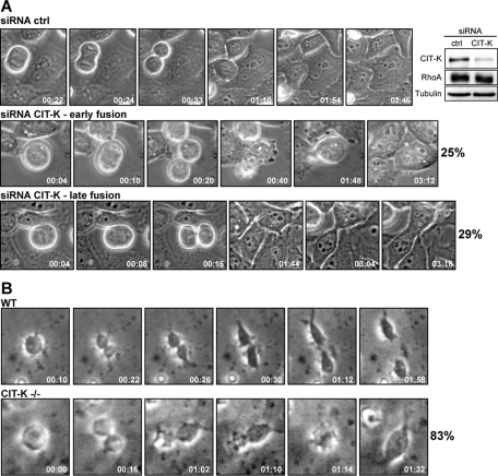
FIGURE 1:
CIT-K is require for abscission both in vitro and in vivo. (A) Selected frames from time-lapse series of HeLa cells undergoing cytokinesis. HeLa cells were transfected with control or CIT-K siRNA and observed 30 h posttransfection by time-lapse video microscopy. Top, A control cytokinesis. CIT-K-depleted cells displayed an “early fusion” (middle) and a “late fusion” (bottom) phenotype. At least 50 cells in cytokinesis were analyzed for each condition. For full movies, see Movies S1–S3. The level of endogenous CIT-K and RhoA expression in whole-cell lysates was determined by immunoblotting using the corresponding antibodies. Tubulin was the internal loading control (right). (B) Selected frames from time-lapse series of GPCs undergoing cytokinesis. GPCs were isolated from P5 wild-type (WT) and CIT-K knockout (CIT-K−/−) mice and observed by time-lapse video microscopy 3 h after plating. At least 40 cells in cytokinesis were analyzed for CIT-K−/−mice. For full movies, see Movies S6 and S7.

