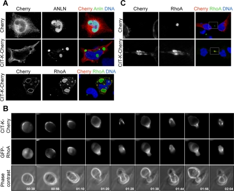
FIGURE 5:
CIT-K overexpression affects the localization of anillin and of active RhoA. (A) HeLa cells were transfected with either Cherry or Cherry-CIT-K. Interphase cells were analyzed by immunofluorescence for localization of the endogenous anillin or active RhoA. (B) Selected frames from time-lapse series of HeLa cells undergoing cytokinesis. HeLa cells were cotransfected with GFP-ceRhoA and CIT-K-Cherry and observed 30 h posttransfection by time-lapse video microscopy. For full movie, see Movie S11. (C) HeLa cells in late cytokinesis, expressing either Cherry or CIT-K-Cherry, were analyzed for the localization of active RhoA by immunofluorescence 30 h posttransfection. Nuclei were counterstained with DAPI.

