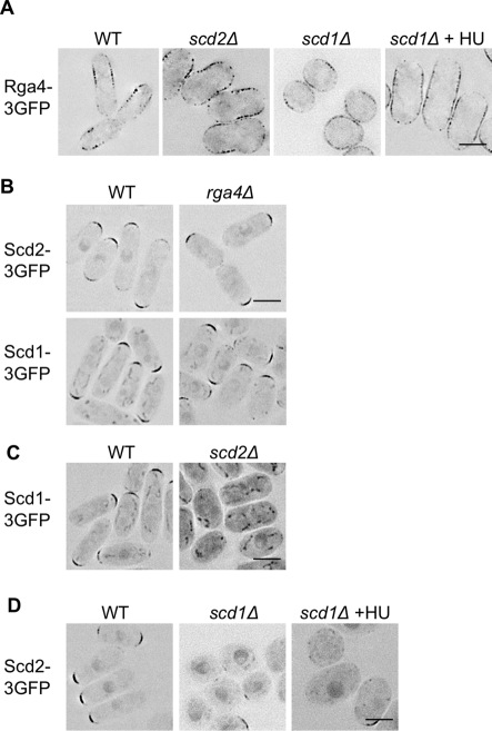
FIGURE 4:
Both pathways can localize independently. (A) Rga4-3×GFP does not require Scd1 or Scd2 for localization to cell sides. (B) Neither Scd1-3×GFP nor Scd2-3×GFP is dependent on Rga4 for localization to cell tips. (C) Scd1-3×GFP is lost from the cell tips in scd2Δ. (D) Scd2-3×GFP is mislocalized in scd1Δ. Images for all are representative, deconvolved, single focal planes with inverted LUTs. Scale bars: 5 μm. Unless marked, cells were in exponential growth at the time of imaging. scd1Δ is also shown after growth for 5 h in HU to elongate the cells.

